Photos & Graphics
Photos

Human Neutrophil Granulocyte
3D Reconstruction of a Human Neutrophil Granulocyte. The image was taken using confocal laser scanning microscopy.


ISAS Executive Board
Prof. Dr. Albert Sickmann (Chairman, photo: left) and Jürgen Bethke (Chief Financial Officer)

Myocardial Infarction: Immune Cell Infiltration
Light sheet fluorescence microscopy of immune cell infiltration (red-orange) in a murine heart (blue) after myocardial infarction. The image was taken using confocal laser scanning microscopy.
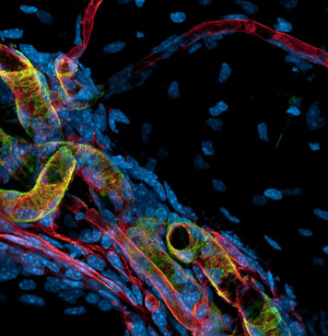
Periosteum: Blood Vessels
To ensure bone homeostasis, the bone surface – the periosteum – and the cortical bone are supplied by different types of blood vessels, such as fine capillaries (red) and large arteries (green and red). The image was taken using confocal laser scanning microscopy.


Rheumatoid Arthritis: Murine Knee Joint (I)
Infiltration of immune cells (red) and inflammation-driven angiogenesis of blood vessels (green and white) in a murine knee joint during rheumatoid arthritis. The image was taken using confocal laser scanning microscopy.

Rheumatoid Arthritis: Murine Knee Joint (II)
Infiltration of immune cells (red) and inflammation-driven angiogenesis of blood vessels (green and white) in a murine knee joint during rheumatoid arthritis. The image was taken using confocal laser scanning microscopy.
Graphics
-
Microscopy Images: Cell tracking
Download fileA visual representation of the trajectory of cells in space over time. Each coloured trajectory stands for a different cell. The x and y axes denote the spatial position of the cells in an image. More information about this graphic via research project “Cell Tracking in Microscopy Images”.
-
NephrESAthrombo
Download fileThe main objectives of NephrESA are developing assays for plasma risk factors for thrombovascular events and for platelet activation status in CDK patients; analysing changes in the dynamics of platelet activation in CKD patients; developing a computer model (NephrESAthrombo submodel) of thrombovascular risks for CKD patients.

